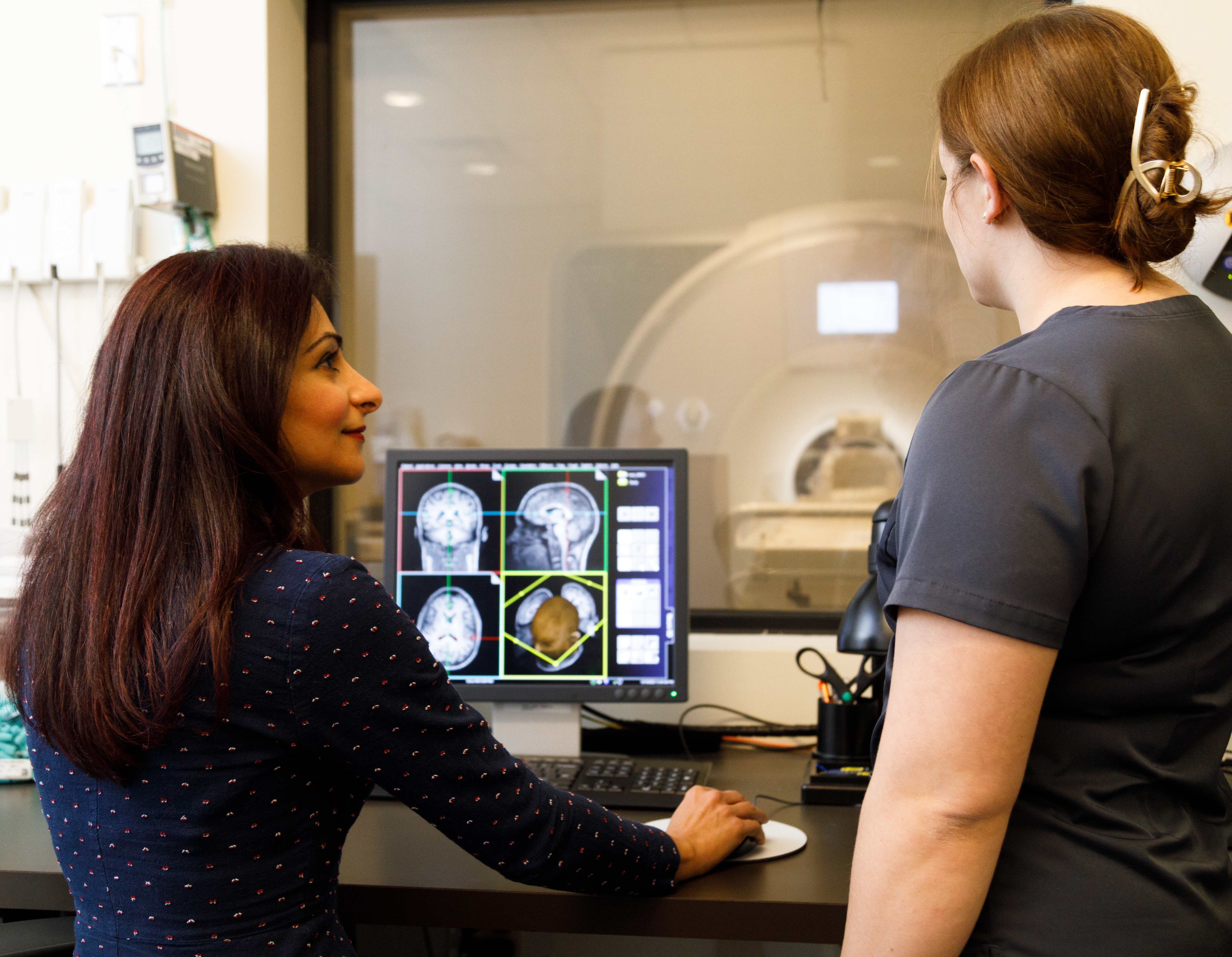
The Foundation is one of the few independent rehabilitation research organizations in the U.S. with a state-of-the-art, research-dedicated MRI scanner. The Ortenzio Center provides Foundation scientists with cutting-edge technology, enabling the development of innovative interventions and delivering imaging capabilities that far exceed those of standard clinical MRI systems.
The Ortenzio Center’s powerful MRI scanner enables scientists to work in real-time, correlating cognitive and motor function with activity in the brain and spinal cord and documenting the effectiveness of new interventions. These findings lead to breakthroughs that enhance recovery and improve outcomes for people with disabilities.

Ortenzio Center Leadership


Advanced multi-modality biomedical techniques allow our researchers to visualize brain activity during specific tasks and sensations in individuals with MS, traumatic brain injury, and Parkinson's disease. Some of the techniques used include functional magnetic resonance imaging (fMRI), electroencephalography (EEG), and electromyography (EMG). Their goal is to understand how cognitive rehabilitation can reshape memory-related brain regions and provide valuable insights into addressing cognitive and physical fatigue.
The Ortenzio Center's leading-edge Siemens 3T scanner enables our scientists to investigate brain mechanisms that support effective rehabilitation strategies and interventions through neuroimaging. Researchers analyze the complex brain-body connection by recording real-time responses during specific motor, language, memory, or visual tasks in the scanner and conducting further offline analysis.
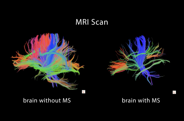
Our researchers are using advanced brain imaging to better understand cognitive fatigue in people with multiple sclerosis. Their work has identified a measurable brain marker that helps evaluate the effectiveness of MS treatments. Studies also show that fatigue in MS is closely linked to changes in brain activity and thinking performance. The image at left shows an MRI scan comparing a brain without MS to one with MS, highlighting structural differences associated with the disease.
Using MRI, our researchers aim to understand how different exercise programs combined with memory retraining can improve cognitive performance in individuals with traumatic brain injury. Functional magnetic resonance imaging scans taken before and after interventions help explain why certain performance changes happen and how differences between exercise groups develop over time.
Several neurobehavioral reading interventions have been developed using functional magnetic resonance imaging and neurofeedback to boost post-stroke brain plasticity. Since reading impairments frequently affect many left-hemisphere stroke survivors, the goal is to target chronic conditions like aphasia through early interventions, enhancing outcomes for those with reading disorders after a stroke.
Ortenzio Center Team


Establishing a Clearer Measure of Mental Fatigue in Multiple Sclerosis
This study examines mental fatigue that is regularly experienced by people with multiple sclerosis. Researchers will examine levels of mental fatigue by looking at brain activation during an MRI scan and analyzing self-reported mental fatigue provided during study participation.
Frequently Asked Questions About MRI and fMRI
Magnetic resonance imaging and functional magnetic resonance imaging are safe, noninvasive methods of producing detailed images of the body and brain without radiation. Most people can undergo these scans.
Magnetic resonance imaging (MRI) is a noninvasive, painless imaging technique that uses a strong magnetic field and radio waves to generate detailed images of organs and tissues in the body. Most MRI machines are large, cylindrical magnets that create a strong magnetic field to interact with hydrogen atoms in the body. This, combined with radiofrequency pulses, produces detailed cross-sectional images or "slices" of anatomy. MRI can also generate 3D images that provide a comprehensive view from multiple angles. At Kessler Foundation, MRI is frequently used in clinical trials to monitor the effect of interventions.
Functional magnetic resonance imaging (fMRI) is a type of MRI that helps assess brain function by measuring and mapping brain activity. A most common fMRI method, called BOLD fMRI, works by detecting changes in blood flow and oxygen levels in the brain that happen when certain areas become more active. When a brain region is in use, it needs more oxygen, which increases blood flow to that area. By capturing these changes, fMRI shows which parts of the brain are active during specific tasks or activities.
An fMRI shows patterns of brain activity, helping researchers understand how different parts of the brain handle things like movement, language, and memory and cognition. This technique has been key in identifying areas involved in thinking and movement, mapping brain connections, and studying changes in brain activity in conditions like stroke, brain injury, and diseases that affect the brain.
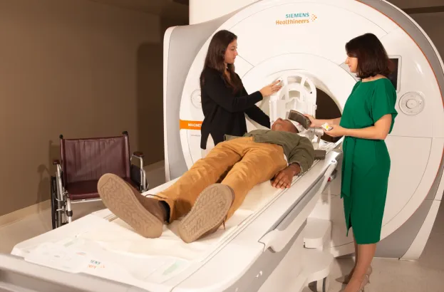
First, you’ll lie down on the MRI table, typically on your back, and the table will slide into the MRI machine’s cylindrical chamber. To ensure clear images, you’ll need to remain as still as possible throughout the scan, which can last between 30 and 90 minutes, depending on the area being imaged and the study task. You may be asked to perform specific tasks or respond to stimuli, such as reading text or moving body parts, while inside the scanner.
You won’t need to undress, but you will be asked to wear clothing without zippers or snaps, like sweatpants and a t-shirt. You’ll also need to remove any metal items, including jewelry, glasses, underwire bras, and belts.
No contrast dyes or IV injections are used in Kessler Foundation MRI or fMRI studies.
MRI is a safe imaging method because it doesn’t use the ionizing radiation found in x-rays and computed tomography (CT) scans. This makes MRI ideal for cases where frequent imaging is needed, especially for brain-related diagnoses and treatments.
Depending on the specific study, scans usually take from 30 to 90 minutes depending on the size of the area being scanned and the number of images being taken.
Having metal in your body doesn’t automatically rule out an MRI scan, but it’s essential to inform the research team. They will ask about your medical history to determine if an MRI is safe for you. In some cases, an MRI may not be recommended. For instance, people with certain implants, especially those containing iron—like pacemakers, nerve stimulators, defibrillators, insulin pumps, cochlear implants, brain stimulators, or capsule endoscopy devices—should avoid MRI machines.
An EEG records brain wave activity, providing detailed insights into brain patterns during neuroimaging. In this painless test, small sensors (electrodes) are gently attached to the scalp with adhesive or an elastic cap. These electrodes capture the brain's electrical activity, which is transmitted via wires to a device that amplifies and records brain waves on a computer.
If you experience claustrophobia, inform the researchers immediately. The Ortenzio Center features a spacious scanner and offers two-way communication between researchers and you, which may ease your claustrophobia. Additionally, the Foundation has a mock MRI scanner that can help you acclimate to the machine, its procedures, and the sounds it produces. To further reduce discomfort, visualization techniques are available, along with supportive options such as listening to music, watching videos, using eye covers, or holding a panic button for reassurance.
For Kessler Foundation studies, we recommend avoiding tranquilizers and certain mood-altering medications or substances, as they can affect brain activity and potentially influence scan results. Researchers will discuss your medications before you take part in a study.
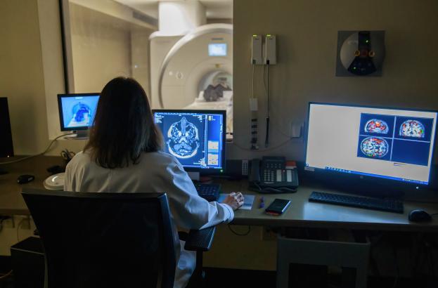
Specialized Laboratory Advancing Neuroimaging Research
